

3‑D Digital Imaging X‑Ray Analysis: Precision Diagnostics for Better Spinal Health
In modern chiropractic care, accurately understanding a patient’s spinal structure is one of the most important steps in delivering individualized, effective treatment. Today’s advanced imaging technologies — especially 3‑D digital imaging X‑ray analysis — give chiropractors a level of detail and precision far beyond what traditional X‑rays or physical exams alone can provide. These tools allow practitioners to see the spine and surrounding skeletal system from multiple angles, measure alignment abnormalities with greater accuracy, and create more precise, evidence‑based care plans.
At Denver Upper Cervical Chiropractic, 3‑D digital imaging X‑ray analysis plays a central role in evaluating the upper cervical spine — the top two vertebrae in the neck — and determining the best course of treatment. This technology improves diagnosis, enhances treatment planning, reduces guesswork, and supports measurable outcomes for patients seeking relief from pain, stiffness, nerve dysfunction, and other musculoskeletal problems.
In this in‑depth article, we’ll explore what 3‑D digital imaging X‑ray analysis really is, how it works, why it’s important in chiropractic care, and the benefits it offers both patients and practitioners.
What Is 3‑D Digital Imaging X‑Ray Analysis?
3‑D digital imaging X‑ray analysis refers to advanced radiographic imaging technology that captures three‑dimensional images of spinal structures, bones, and joints. Unlike traditional 2‑D X‑rays, which provide flat images in one or two planes, 3‑D imaging allows clinicians to view the anatomy from multiple angles and depths, making structural abnormalities and misalignments easier to identify.
Within upper cervical chiropractic (the specialty practiced at Denver Upper Cervical Chiropractic), 3‑D digital imaging is especially valuable because the atlas (C1) and axis (C2) — the top two vertebrae — have a complex range of motion and play a crucial role in nervous system communication. Misalignment in this region may not be clearly visible on standard X‑rays, but 3‑D imaging helps reveal subtle deviations that can influence symptoms throughout the body.
Why 3‑D Imaging Is More Advanced Than Standard X‑Rays
Traditional X‑rays have long been used in chiropractic to visualize bone structure, assess alignment, and spot fractures or significant structural changes. However, they have limitations:
- They produce flat, two‑dimensional images, which can obscure overlapping structures.
- Anatomical distortions are common, especially in complex regions like the cervical spine.
- Diagnoses based on 2‑D imaging may miss subtle misalignments or rotations.
By contrast, 3‑D digital imaging X‑ray analysis — such as cone‑beam computed tomography (CBCT) — captures a full volumetric representation of structures, allowing clinicians to view the spine from all directions and measure angles and asymmetries with high precision. This leads to better diagnostics and more targeted care.
In upper cervical care, this means that chiropractors can identify not just whether the C1 or C2 vertebrae are misaligned, but exactly how they deviated (e.g., rotated, tilted, or translated) — information that is critical for precise correction.
How 3‑D Digital X‑Ray Analysis Works
The process of 3‑D digital imaging analysis typically involves the following steps:
1. Image Capture
Using advanced digital X‑ray machines or Cone‑Beam CT (CBCT) technology, the patient is scanned in a manner that produces a three‑dimensional data set. This can be done while the patient is seated, standing, or in a specific head/neck position depending on the target area.
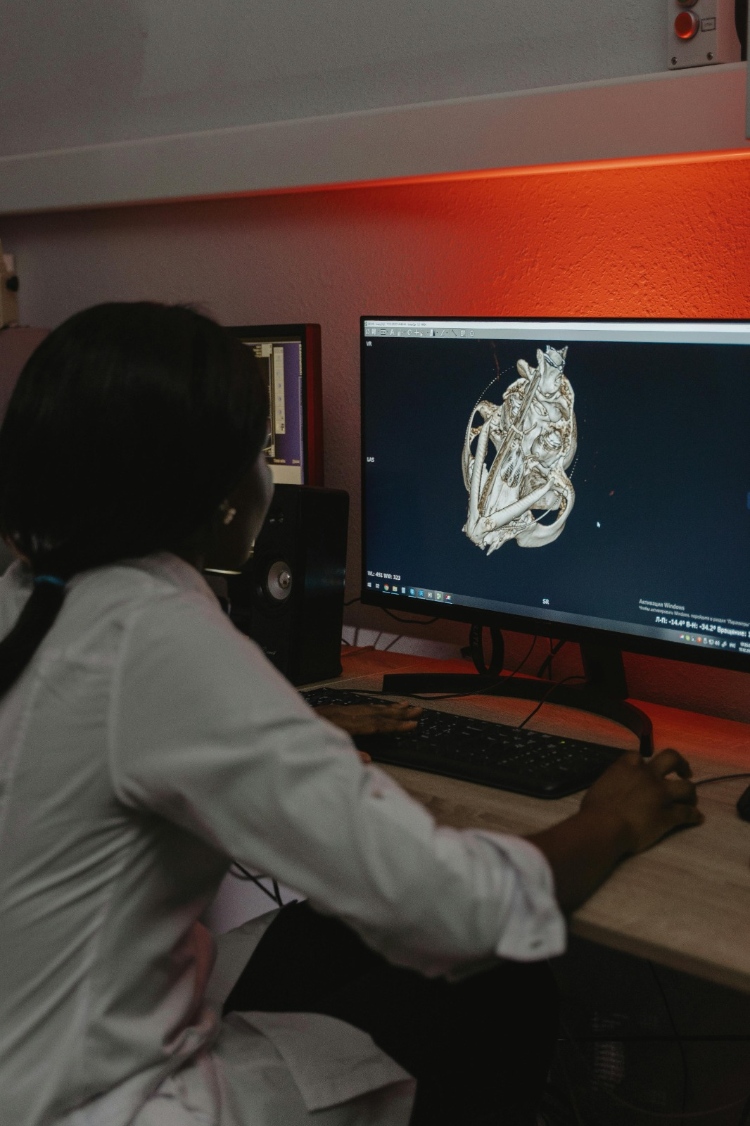
2. Digital Reconstruction
The captured radiographs are digitally reconstructed using specialized software that creates a detailed 3‑D model of the area. This can be rotated, zoomed, and viewed from multiple angles to reveal structural relationships that would not be visible on a flat X‑ray.
3. Analysis and Measurement
From the 3‑D model, clinicians can measure:
- Vertebral rotation
- Joint spacing
- Asymmetry
- Structural deviations
- Alignment relative to the central axis
This level of detail helps chiropractors plan precise adjustments tailored to the patient’s unique anatomy.
4. Treatment Planning
After review, the chiropractor uses the 3‑D information to develop a targeted care plan that directs adjustment vectors and therapeutic strategies with millimeter‑level accuracy.
The Role of 3‑D Digital Imaging X‑Ray Analysis in Upper Cervical Chiropractic
Upper cervical chiropractic focuses on the first two cervical vertebrae (atlas and axis) because of their crucial roles in posture, balance, head movement, and nervous system communication. Misalignment of these vertebrae can affect how the spinal cord, brainstem, and surrounding nerve pathways function, potentially contributing to pain, headaches, dizziness, and other systemic symptoms.
3‑D digital imaging analysis allows chiropractors like Dr. Ty Carzoli at Denver Upper Cervical Chiropractic to accurately detect misalignment patterns and their impact on the neural canal. Through this sophisticated imaging, adjustments can be delivered precisely, using minimal force and maximizing the chance of long‑lasting correction.

Benefits of 3‑D Digital X‑Ray Analysis in Chiropractic Care
There are several key advantages that 3‑D digital imaging X‑ray analysis offers over traditional techniques:
1. Enhanced Diagnostic Clarity
3‑D imaging eliminates the overlap and distortions common in 2‑D X‑rays, allowing practitioners to see deeper and clearer into the spine’s structure. This clarity improves the ability to detect subtle misalignments, joint degeneration, or congenital abnormalities that could affect treatment outcomes.
2. Precision Measurement for Precise Adjustments
Because upper cervical adjustments often rely on tiny changes in vertebrae position, knowing the exact degree and direction of misalignment is essential. 3‑D imaging provides these measurements with high accuracy, enabling chiropractors to design instrument‑assisted adjustments that target the precise vector needed for correction.
3. Customized Treatment Plans
By understanding a patient’s unique anatomy, chiropractors can tailor care plans instead of relying on generalized adjustment techniques. This increases the likelihood of better, longer‑lasting outcomes and can reduce the number of visits needed.
4. Better Patient Education
Seeing one’s own 3‑D spinal images makes it easier for patients to understand the source of their symptoms and engage more actively in treatment. Digital images can be annotated, zoomed, and rotated for clearer communication.
5. Reduced Radiation Exposure
Compared to multiple traditional X‑ray views, modern 3‑D imaging systems like CBCT often expose patients to less radiation while still capturing significantly more information.
6. Tracking Progress Over Time
Baseline 3‑D images can be compared to future scans, allowing chiropractors to monitor structural changes, track improvement, and adjust care plans as needed.
How 3‑D Imaging Supports Patient Care
Beyond the technical benefits, 3‑D digital imaging improves the entire patient care experience:
More Informed Decisions
Without advanced imaging, chiropractors may have to infer underlying structural issues based on palpation and symptoms alone. 3‑D analysis gives concrete visual evidence that enhances confidence in diagnosis and treatment selection.
Communication and Transparency
Patients can view their own images directly with the chiropractor, which builds trust and understanding of how their body is structured and what the treatment aims to accomplish.
Coordination With Other Providers
If a patient’s condition requires referral to another provider (e.g., for an MRI or surgical consultation), 3‑D imaging makes it easier to share detailed diagnostics quickly and securely.
Common Uses of 3‑D Digital Imaging X‑Ray Analysis in Practice
While every chiropractic practice is different, the technology is especially helpful in cases where:
- Upper cervical misalignment is suspected
- Past injuries (like car accidents or falls) may have caused subtle structural changes
- Symptoms don’t respond to initial care
- Nerve‑related symptoms (e.g., radiculopathy, tingling, numbness) are present
- Precise adjustment vectors are needed to avoid unnecessary force
Safety and Radiation Concerns
A common question patients ask is about radiation exposure from imaging. Fortunately, digital imaging systems — and specifically CBCT — are designed to use less radiation than traditional CT scans while still providing rich diagnostic detail. This balance of safety and clarity makes 3‑D imaging a valuable tool in chiropractic, particularly for spinal analysis.
Limitations and Considerations
While 3‑D digital imaging X‑ray analysis is powerful, it isn’t necessary for every chiropractic visit or every case. Clinical guidelines recommend imaging when the information will clearly influence the diagnosis or treatment plan, rather than as a routine for all patients.
It’s also vital that imaging is interpreted by trained professionals, because raw images may be misleading without appropriate clinical context. This is why chiropractors who use 3‑D imaging often undergo additional training to ensure that diagnostic data truly informs care.
Conclusion: The Future of Chiropractic Diagnostics
3‑D digital imaging X‑ray analysis is transforming how chiropractors diagnose and treat spinal conditions. It represents a shift from traditional, flat 2‑D imaging to a more holistic understanding of the body’s three‑dimensional structure. For upper cervical chiropractors — who focus on the delicate and nuanced alignment of the atlas and axis — this technology is especially impactful, helping clinicians detect hidden misalignments, tailor precise adjustments, and track progress with clarity and confidence.
By leveraging advanced digital imaging, practices like Denver Upper Cervical Chiropractic are able to offer more accurate diagnoses, better patient education, and customized, effective care that supports long‑term wellness. Whether you’re experiencing chronic pain, post‑injury symptoms, or unexplained neurological issues, 3‑D digital imaging offers a diagnostic edge that enhances treatment outcomes and empowers patients to take control of their spinal health.
© 2026 Denver Upper Cervical Chiropractic